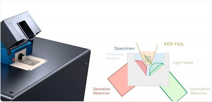
The androgen receptor (AR) is a nuclear receptor that plays a role in development and maintenance of male phenotype. Malfunctioning of AR is closely associated with prostate cancer, the most prominent form of cancer in males. Upon activation by testosterone, AR translocates to the nucleus, where it binds to DNA and induces transcription of specific genes. During this project you will study the dynamics and location of the AR. For this you will make use of cells expressing fluorescently tagged AR proteins and a Luxendo lightsheet microscope (InviSPIM). In lightsheet microscopy the sample is illuminated through an extra lens perpendicular to the lens used for image acquisition. Using this microscope we can selectively illuminate a sheet in the sample, and image the complete field of view very fast using a sensitive sCMOS camera. This allows us to monitor dynamic proteins within the 3D environment of the cell.
In this project you will learn how to setup and optimize the complete imaging experiment, from sample preparation to live cell imaging and subsequent image analysis. The InviSPIM is equipped with a spatial light modulator (SMD) to create different shapes of the illuminating sheet. Optimizing the beam shape for different kind of imaging experiments will be useful for the future users of this microscope. This study will lead to better understanding of the functioning of AR in 3-D cultured cells, including AR receptor dimerization and its DNA binding properties.

Contact Information:
Erasmus Optical Imaging Center
www.erasmusoic.nl
Johan Slotman (j.slotman [a] erasmusmc.nl)
Tel: 010-7037644

