

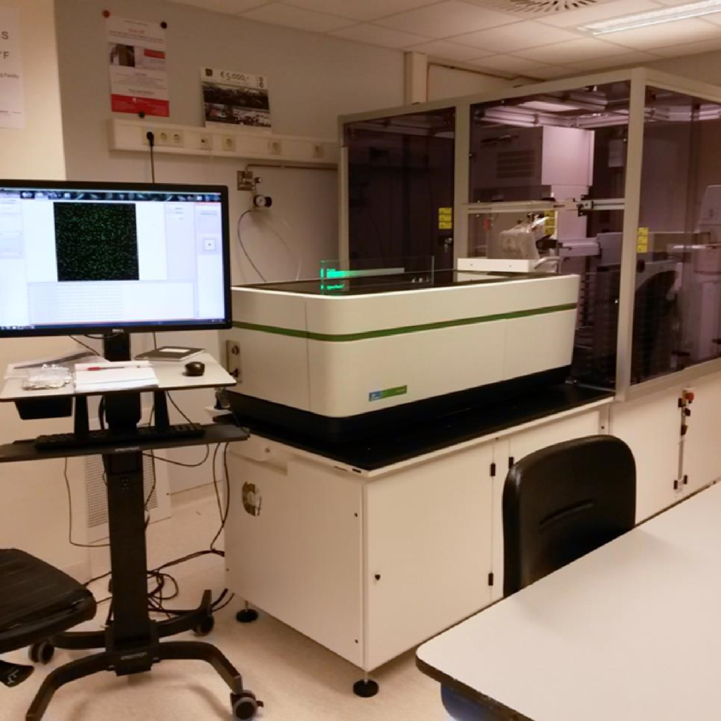
High Content Screening System with automation and liquid handling, Perkin Elmer
Lasers

Objectives
| Magnification | Numerical Aperture | Working Distance (mm) | Immersion | Type |
| 5x | 0.16 | long WD | Air | |
| 10x | 0.3 | long WD | Air | |
| 20x | 0.4 | 8.28 | Air | |
| 40x | 1.1 | Water |
Camera
2x sCMOS camera (4.4 megapixels, 6.5 μm x 6.5 μm pixel size)
Sample Holders
Multi-well (96/384) plates, up to 4 slides simultaneously
(Other sample types can be discussed)
Automation and liquid handling
Robotics: Plate handler workstation SX 2
Incubator: 40 multi-well plate incubator (37°C, 5% CO2, 95% humidity)
Dispensor: Janus 4-tip
Washer: ELx405 (50 – 3000 μL/well)
Barcode reader
Extra info
System contains both Spinning disk confocal and Widefield fluorescence optics.
Spinning disk module with Confocal SynchronyTM Optics (Perkin Elmer).
Imaging modes: Fluorescence, brightfield and digital phase contrast
Available band pass filters: 435-480, 435-515, 435-550, 500-550, 570-630 and 650-760 nm
Available analysis software
Harmony® High Content Imaging and Analysis Software provides a complete solution to set up assays and automate your high content imaging experiments, acquire images and analyze data, and then store, retrieve and present the results.
The PhenoLOGIC™ machine-learning option enables our High Content Analysis software to recognize different cell populations and regions using a simple learn-by-example approach. PhenoLOGIC sets parameters for optimal image segmentation and cell classification. The software combines the most meaningful parameters, whether it’s two, three, four or more, to achieve accurate classification of cells. The result is highly robust and statistically relevant results.
Columbus™ Scope system is a universal high-volume image data storage and analysis system that rings access to images from a wide range of sources including all major high content screening instruments via the Internet.
ImageJ/Fiji is a public domain Java image processing and analysis package. Customized image analysis can be performed in collaboration with Erasmus Optical Imaging Centre (OIC) and Biomedical Imaging Group Rotterdam (BIGR).