Categories
Imaging Facility OIC News

Merry Christmas and a happy 2026!

The Erasmus Optical Imaging Centre wishes you a happy 2026!!!

Tsion, Martijn, Marla, Johan, Jeffrey, Ilse, Gert-Jan, Bart, Alex and Adriaan

Confocal image of a live neuronal growth cone searching for a fellow 
neuron to make synapses together. Regulatory protein ABI1 (red) 
is present at the tips of growing actin filaments (green) as well as at the 
front edges of the lamellae connecting the filaments.
Image recorded by Tim Allertz & Jeffrey van Haren, OIC.

Categories
Course Imaging Facility OIC News

OIC COURSE “Microscopic Image Analysis: From Theory to Practice”

On Thursday and Friday July 3rd-4th 2025 the OIC organizes, in cooperation with the ErasmusMC Graduate School a two-day Image analysis course “Microscopic Image Analysis: From Theory to Practice”. This course focuses on first time users and explains the basis of image analysis and how to use this in Fiji (ImageJ).

Course description 

Microscopic images contain much more information than normally is retrieved. Extracting this information by visual inspection and manual measurement is not only cumbersome but also subjective. Automation of image analysis tasks by using a computer and the right software tools allows for higher efficiency, accuracy, objectivity, reproducibility, and completeness. 

Requirements 

Participants should bring their own laptop to the course for the practical exercises. Software and sample image data will be provided during the course. No prior knowledge of image processing is required. Participants have the opportunity to bring their own image data and directly apply the newly acquired image analysis techniques. 

Link to registration: Microscopic Image Analysis: From Theory to Practice

Categories
Demo Imaging Facility Microscope

Abbelight TIRF MN360 Demo

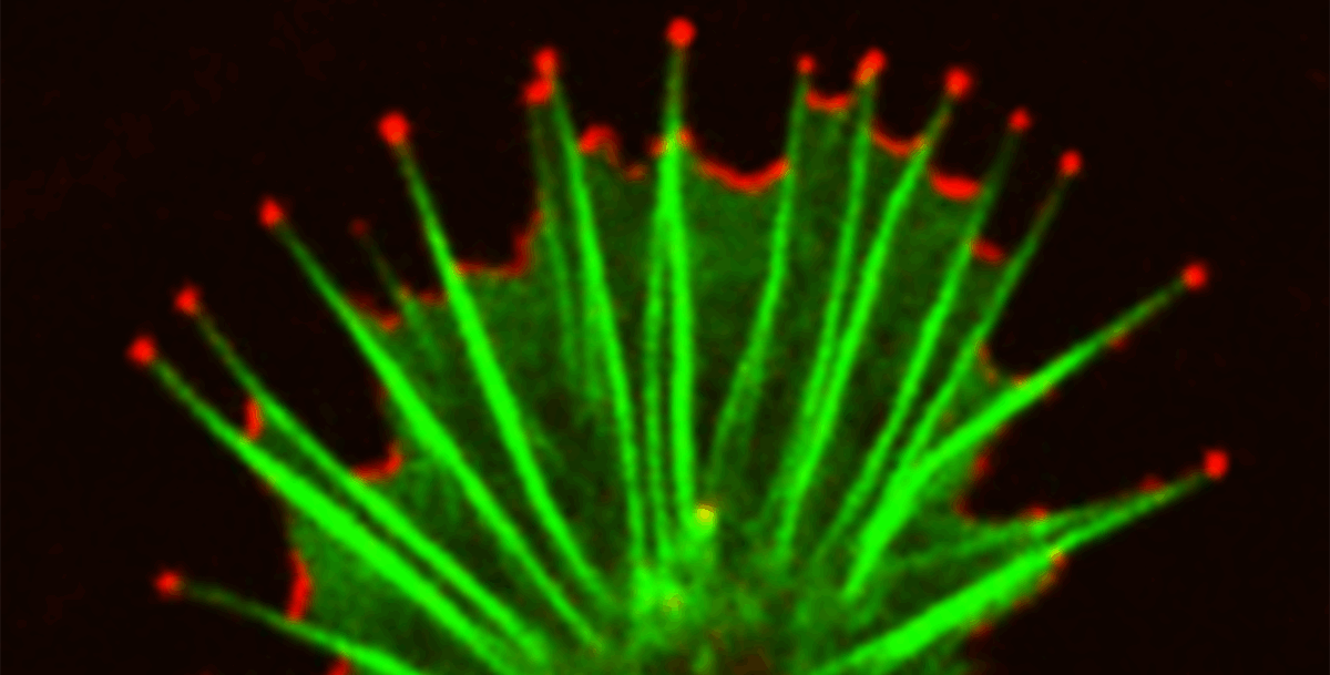
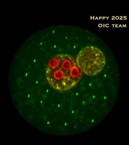
Multicolor, Single molecule localization (dSTORM, PALM, and PAINT) and TIRF microscopy

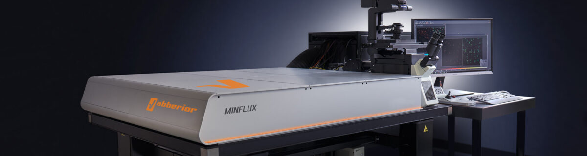
We are excited to announce that our imaging facility has a state-of-the-art Abbelight MN360 system available for demonstation from July 7 till 11.

The Erasmus Optical Imaging Centre together with Abbelight will host a SAFe MN630 demonstration. This add on module is used for multicolor Total Internal Reflection Fluorescence (TIRF) that exploits the unique properties of an induced evanescent wave in a limited specimen region immediately adjacent to the interface between two media having different refractive indices (e.g.: the contact area between a specimen and a glass coverslip).

The MN360 offers an optimal and simultaneous multicolor imaging with the benefits of ASTER technology. This technology provides an ultra-widefield and speckle-free illumination of biological structures close to the coverslip such as membranes, focal adhesions, and much more.  

For more information on the system visite the Evident website: https://evidentscientific.com/en/products/super-resolution/abbelight

For more information on the demonstration please contact Gert-Jan Kremers

Categories
Imaging Facility OIC News

Update: Microscope calendar improvements

The microscope booking calendar has been updated with a few helpful new features. These changes make the interface more flexible and easier to use.

The calendar now supports both light and dark mode, with colors adjusted for clear visibility in each. In the month view, a new “Day” button allows users to quickly switch to the detailed daily view.

If you have any questions or feedback, feel free to reach out to the Erasmus OIC team.

Categories
Imaging Facility OIC News

Happy 2025

The Erasmus Optical Imaging Centre wishes you a happy 2025!!!

Tsion, Martijn, Johan,
Marla, Bart, Ilse, Gert-Jan, Alex,
Adriaan & Gert

Confocal image (circular crop) of a fertilized mouse oocyte, with male and female pronuclei (yellow) and four prominent nucleoli (red) in the female pronucleus. The oocyte was decorated with ‘candle lights’ which are copies from the rightmost bright tiny structure in the cytoplasm (original image recorded by Gert van Cappellen, OIC)

Categories
Course Demo Imaging Facility OIC News

MinFlux super-resolution
demo & workshop

The Erasmus Optical Imaging Centre will host a MinFlux demonstration in the period of November 11-15 and a NVvM workshop focussed on MinFlux microscopy on Monday November 18. 

MinFlux 3D unrivaled resolution and speed

MINFLUX, or minimal fluorescence photon fluxes microscopy, is a super-resolution light microscopy method that offers nanometer-scale 3D-imaging and microsecond-range single molecule tracking and can attain 1–3 nm resolution in three dimensions

We are excited to announce that our imaging facility has a state-of-the-art Abberior MinFlux system available for demonstation November 11-15. The system will also be part of the OIC functional imaging course in the same week.
The demonstration will be concluded with a whole day NVvM MinFlux workshop on Monday November 18, 2024.

More information about the workshop, demonstration period and the registration can be found here.

Categories
Imaging Facility OIC News

Our new colleague Ilse

We would like to introduce our new colleague Ilse Bakker, who started Juny of this year.

Ilse will be working in the OIC in the context of a project financed by the Convergence Flagship Imaging Facility and Innovation Centre (CIFIC) lead by Adriaan Houtsmuller, Erasmus MC and Jacob Hoogeboom, TU Delft. She will develop novel methods and staining techniques for combining fluorescent confocal microscopy and electron microscopy.

Categories
Imaging Facility OIC News

Our new colleague Marla

We would like to introduce our new colleague Marla Lavrijsen, she started this april within the OIC.

She will be a new member of our team assisting users of our confocal microscopes during their experiments and introductions.

Categories
Imaging Facility OIC News

NWO Roadmap NL-BioImaging started

The NWO Roadmap consortium NL-bioimaging has officially started.

The Erasmus Optical Imaging Centre is one of the imaging nodes within NL-BioImaging, an infrastructure for advanced microscopy within the Netherlands. With the the start of this initiative the OIC receives a five year funding for development of automated microscopy.