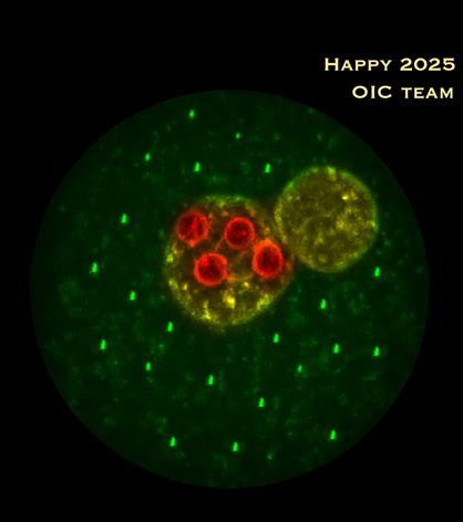
The Erasmus Optical Imaging Centre wishes you a happy 2026!!!
Tsion, Martijn, Marla, Johan, Jeffrey, Ilse, Gert-Jan, Bart, Alex and Adriaan
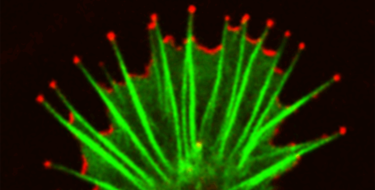
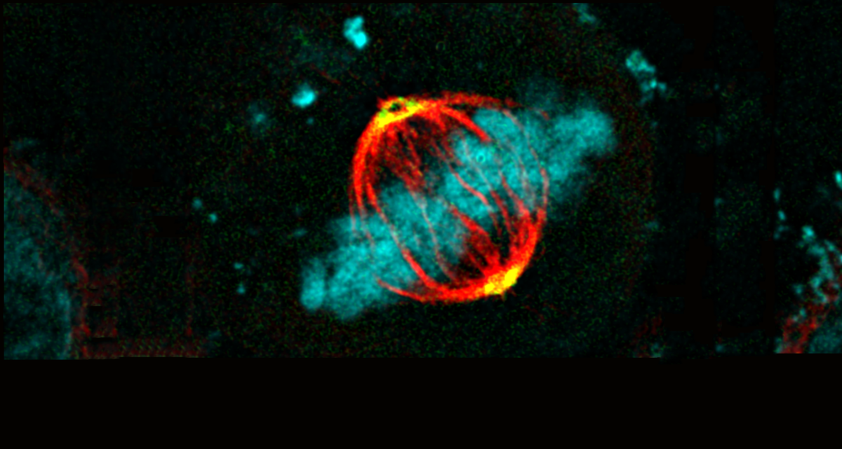
Confocal image of a live neuronal growth cone searching for a fellow neuron to make synapses together. Regulatory protein ABI1 (red) is present at the tips of growing actin filaments (green) as well as at the front edges of the lamellae connecting the filaments. Image recorded by Tim Allertz & Jeffrey van Haren, OIC.
On Thursday November 20th 2025 the OIC and the AMIE facility join forces to highlight the possibilities of cutting edge imaging with research and diagnostic applications.
Location: Ae-406
Program and Speakers
10:00 – 10:10
Opening
10:10 – 10:50
Riëtte de Bruin
Dept. of Otorhinolaryngology and Head and Neck Surgery, Erasmus MC
Title: Photodynamic therapy and optical diagnostics
10:50 – 11:30
Bo Zweedijk
Dept. of Surgery, Erasmus MC
Title: Clinical applications of image-guided surgery
11:30 – 11:50
Break
11:50 – 12:30
Yannick Taverne
Dept. of Cardiothoracic surgery, Erasmus MC
Title: Biofeedback modalities of living myocardial slices
12:30 – 13:10
Matthijs Snelders
Dept. of Molecular Genetics, Erasmus MC
Title: Implementation of advanced microscopy in organ-on-chip devices.
13:10 – 14:20
Lunch (provided) with industry lectures
14:20 – 15:00
Nathaniel Germain
Dept. of Neuroscience, Erasmus MC
Title: The translational value of MRI and non-biased analysis approaches for the understanding of neurodevelopmental disorders.
Keynote speaker
15:00 – 16:00
Massimo Mastrangeli
Dept. of Microelectronics, TU Delft
Title: Microelectromechanical physiological systems for cardiac, neuronal, and tissue barrier applications.
On Thursday and Friday July 3rd-4th 2025 the OIC organizes, in cooperation with the ErasmusMC Graduate School a two-day Image analysis course “Microscopic Image Analysis: From Theory to Practice”. This course focuses on first time users and explains the basis of image analysis and how to use this in Fiji (ImageJ).
Course description
Microscopic images contain much more information than normally is retrieved. Extracting this information by visual inspection and manual measurement is not only cumbersome but also subjective. Automation of image analysis tasks by using a computer and the right software tools allows for higher efficiency, accuracy, objectivity, reproducibility, and completeness.
Requirements
Participants should bring their own laptop to the course for the practical exercises. Software and sample image data will be provided during the course. No prior knowledge of image processing is required. Participants have the opportunity to bring their own image data and directly apply the newly acquired image analysis techniques.
The microscope booking calendar has been updated with a few helpful new features. These changes make the interface more flexible and easier to use.
The calendar now supports both light and dark mode, with colors adjusted for clear visibility in each. In the month view, a new “Day” button allows users to quickly switch to the detailed daily view.
If you have any questions or feedback, feel free to reach out to the Erasmus OIC team.
Confocal image (circular crop) of a fertilized mouse oocyte, with male and female pronuclei (yellow) and four prominent nucleoli (red) in the female pronucleus. The oocyte was decorated with ‘candle lights’ which are copies from the rightmost bright tiny structure in the cytoplasm (original image recorded by Gert van Cappellen, OIC)
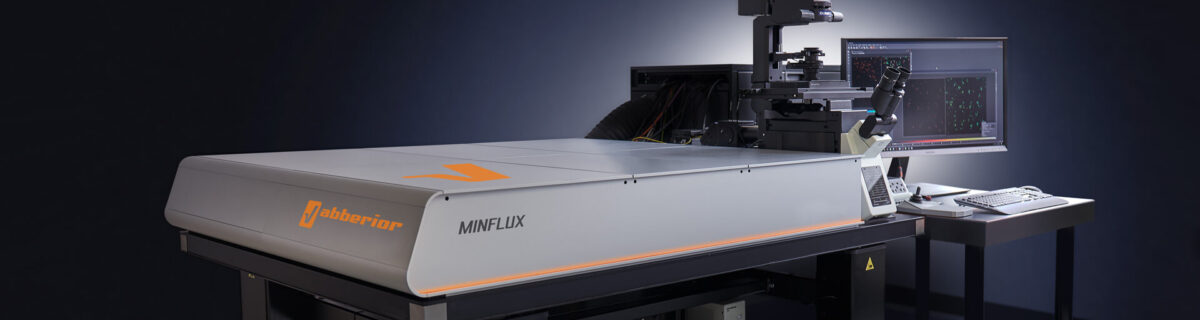
The Erasmus Optical Imaging Centre will host a MinFlux demonstration in the period of November 11-15 and a NVvM workshop focussed on MinFlux microscopy on Monday November 18.
MinFlux 3D unrivaled resolution and speed
MINFLUX, or minimal fluorescence photon fluxes microscopy, is a super-resolution light microscopy method that offers nanometer-scale 3D-imaging and microsecond-range single molecule tracking and can attain 1–3 nm resolution in three dimensions
We are excited to announce that our imaging facility has a state-of-the-art Abberior MinFlux system available for demonstation November 11-15. The system will also be part of the OIC functional imaging course in the same week. The demonstration will be concluded with a whole day NVvM MinFlux workshop on Monday November 18, 2024.
More information about the workshop, demonstration period and the registration can be found here.
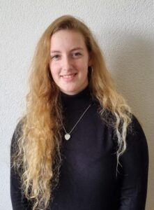
We would like to introduce our new colleague Ilse Bakker, who started Juny of this year.
Ilse will be working in the OIC in the context of a project financed by the Convergence Flagship Imaging Facility and Innovation Centre (CIFIC) lead by Adriaan Houtsmuller, Erasmus MC and Jacob Hoogeboom, TU Delft. She will develop novel methods and staining techniques for combining fluorescent confocal microscopy and electron microscopy.
Registration is not longer possible for our PhD functional imaging and super-resolution course on November 11-15 2024.
The course is full, your registration will be placed on a waiting list. The course will be organized again in the fall of 2025.
This intensive course of one week gives an overview of the rapidly developing advanced fluorescence imaging methods and how they are used in biomedical research in a combination of lectures and hands-on practicals at our microscopes.
More information about the course and the registration form can be found via this link.
On Monday and Tuesday Jan 29th-30th 2024 the OIC organizes in cooperation with the Erasmus MC graduate school the two-day Image analysis course “Microscopic Image Analysis: From Theory to Practice”. This course focuses on first time users and explains the basis of image analysis and how to use this in Fiji (ImageJ).
Course description
Microscopic images contain much more information than normally is retrieved. Extracting this information by visual inspection and manual measurement is not only cumbersome but also subjective. Automation of image analysis tasks by using a computer and the right software tools allows for higher efficiency, accuracy, objectivity, reproducibility, and completeness.
Requirements
Participants should bring their own laptop to the course for the practical exercises. Software and sample image data will be provided during the course. No prior knowledge of image processing is required. Participants have the opportunity to bring their own image data and directly apply the newly acquired image analysis techniques.
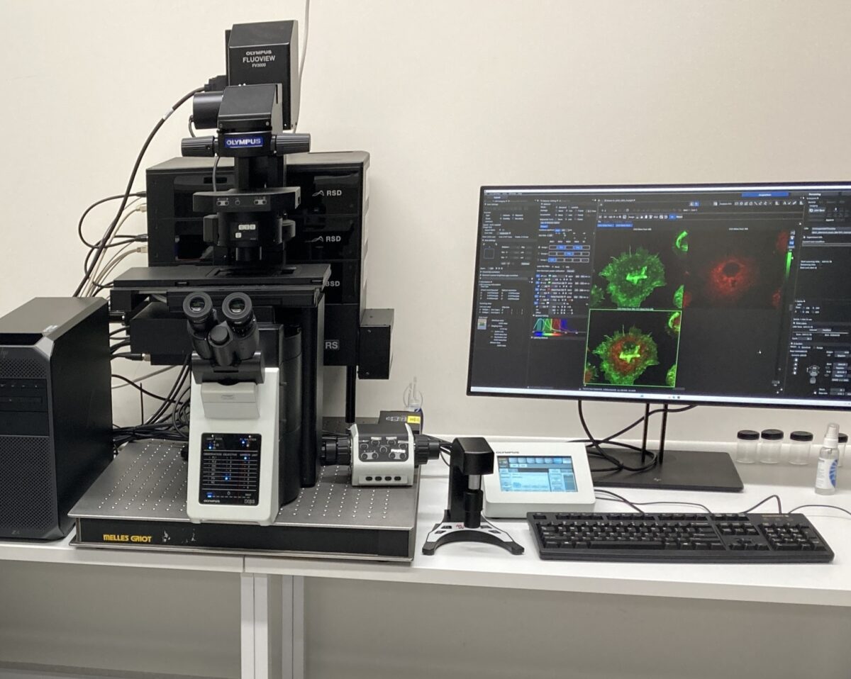
From February 5th to 9th 2024 Evident (formerly Olympus), in association with the OIC, will organize a demonstration of the newly released Fluoview FV4000 point scanning confocal.
Researchers who are interested to test the possibilities of this new generation laser scanning confocal are invited to bring their own samples for testing.
The FV4000 can image most of the regular samples used on the current OIC confocals. In addition, the confocal setup should be especially interesting for researchers using near infrared (NIR) fluorescent probes, or more than 4 fluorescent stains.
Special objectives will be available for imaging organoids and tissues. Specifications of the demonstration setup include:
Inverted microscope
10 laser spanning the wavelength range from 405 nm – to 785 nm
6 detectors for simulaneous imaging and spectral unmixing
Incubation system for live cell imaging (37°C/5% CO2) using 35mm dishes, multiwell slides and multiwell dishes
Resonant scanner for fast (>20fps) imaging
If you are interested to test the FV4000, please send us an email with a short description of the sample you would like to image.