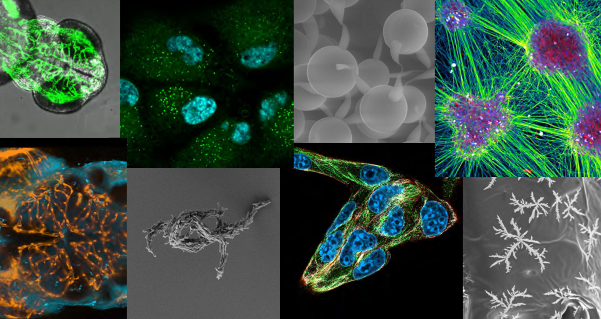
Submit your pictures for the Dutch Microscopy Pictures Calendar 2027. Dig out your best images, spread the word among your colleagues and networks, and feel free to think out of the box!
Deadline to submit: 15 September 2026 The best pictures will be featured on the 2027 Microscopy.nl birthday calendar. For questions email nl-bi@amsterdamumc.nl
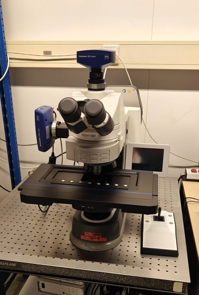
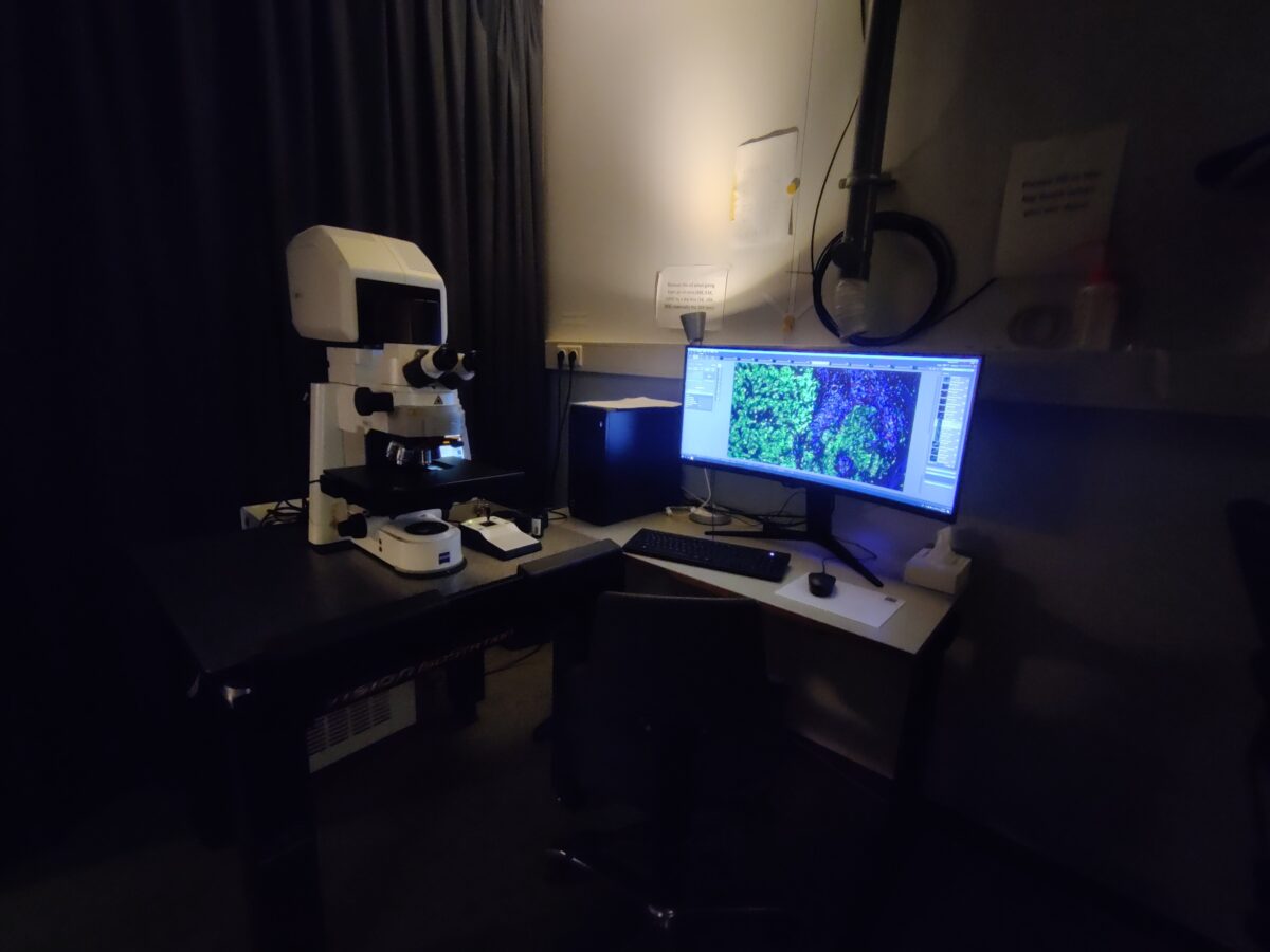
With this news post we would like to introduce the Zeiss AxioImager II Wide-Field microscope which has recently become part of our imaging centre.
This fully motorized system enables fast and scalable overview acquisition of fluorescence images in Wide-Field mode. Compatible with known channels across the visible spectrum and even into the near-infrared range.
An additional color camera allows you to correlate fluorescence images with a brightfield stain such as H&E. The 8‑slide insert makes it possible to process multiple samples in one run, significantly improving throughput for larger experiments.
Previously, this system was part of Pathology’s Research Facility (PARTS). We are pleased to make this microscope available once again within the Optical Imaging Centre. In the near future a software upgrade will make this system ready to be used for SMART microscopy. We will inform you in more detail when this is implemented.
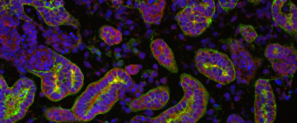
Convenient pre-scan before acquiring confocal images
The microscope is equipped with a motorized stage capable of holding up to eight slides, thereby enabling the setup of large tile scans. The objectives are all air-immersion which makes it possible to adjust the magnification of the objective with the lowest magnification (2.5x) to the highest (63x) magnification without touching your sample.
Although widefield imaging provides lower z‑resolution than confocal microscopy, the system excels in scalability, acquisition speed and low magnification overview imaging. Combining the the two systems will give you an advantage in overview with the widefield and local detailed imaging with the confocal microscope.
The ability to transfer the overview images acquired with the Zeiss AxioImager II microscope to the Zeiss LSM 900 confocal system, also located in Be‑311, and subsequently acquire high‑resolution images of relevant regions, will be of significant value.
More specifications
The extra color camera will make this our only system in the imaging facility to correlate a chromogenic staining like H&E with fluorescence. Additional optics enable polarized light transmission imaging, expanding the range of contrast methods.
The Colibri 7 LED excitation system provides dedicated LEDs for each fluorescence channel, combining long lifetime with rapid switching. Its high transmission in the 735/40 nm band enables efficient near‑infrared imaging with reduced exposure times compared to other systems.
If you want to make use of this microscope, please contact us for an introduction. The system can be found in our schedular under the name WF_311.
More detailed specifications of this system can be found on our equipment page: link.
Multicolor, Single molecule localization (dSTORM, PALM, and PAINT) and TIRF microscopy
We are excited to announce that our imaging facility has a state-of-the-art Abbelight MN360 system available for demonstation from July 7 till 11.
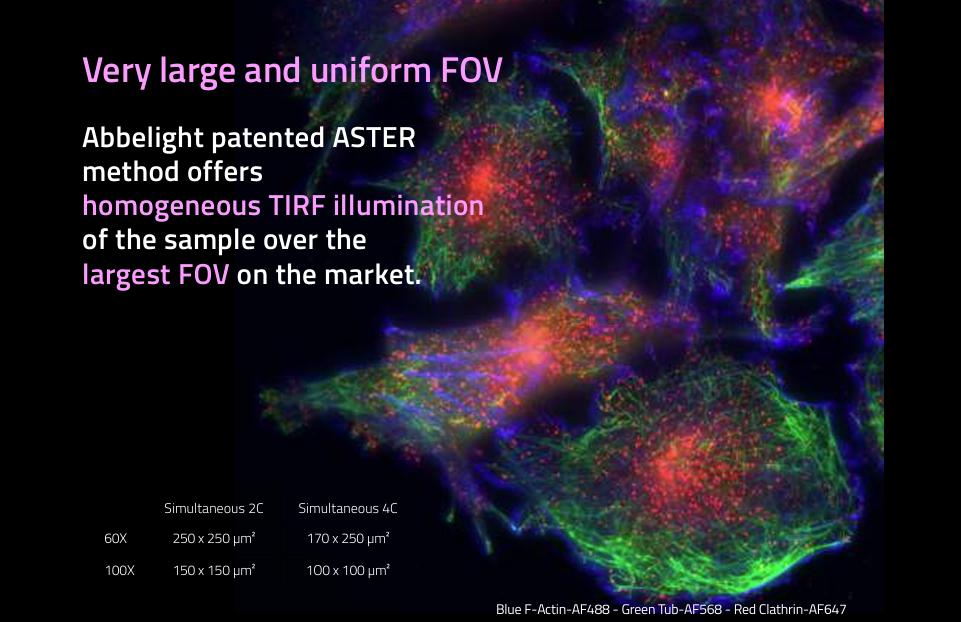
The Erasmus Optical Imaging Centre together with Abbelight will host a SAFe MN630 demonstration. This add on module is used for multicolor Total Internal Reflection Fluorescence (TIRF) that exploits the unique properties of an induced evanescent wave in a limited specimen region immediately adjacent to the interface between two media having different refractive indices (e.g.: the contact area between a specimen and a glass coverslip).
The MN360 offers an optimal and simultaneous multicolor imaging with the benefits of ASTER technology. This technology provides an ultra-widefield and speckle-free illumination of biological structures close to the coverslip such as membranes, focal adhesions, and much more.
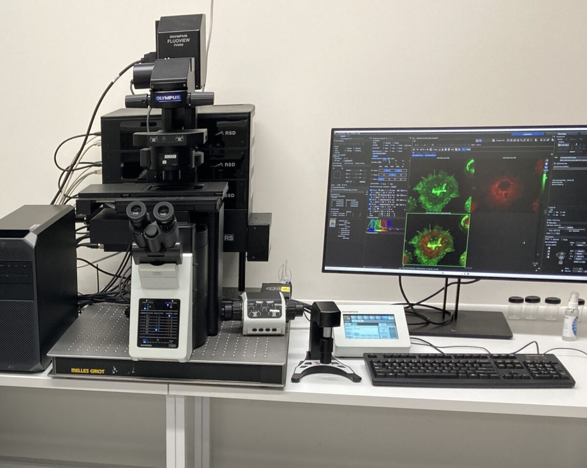
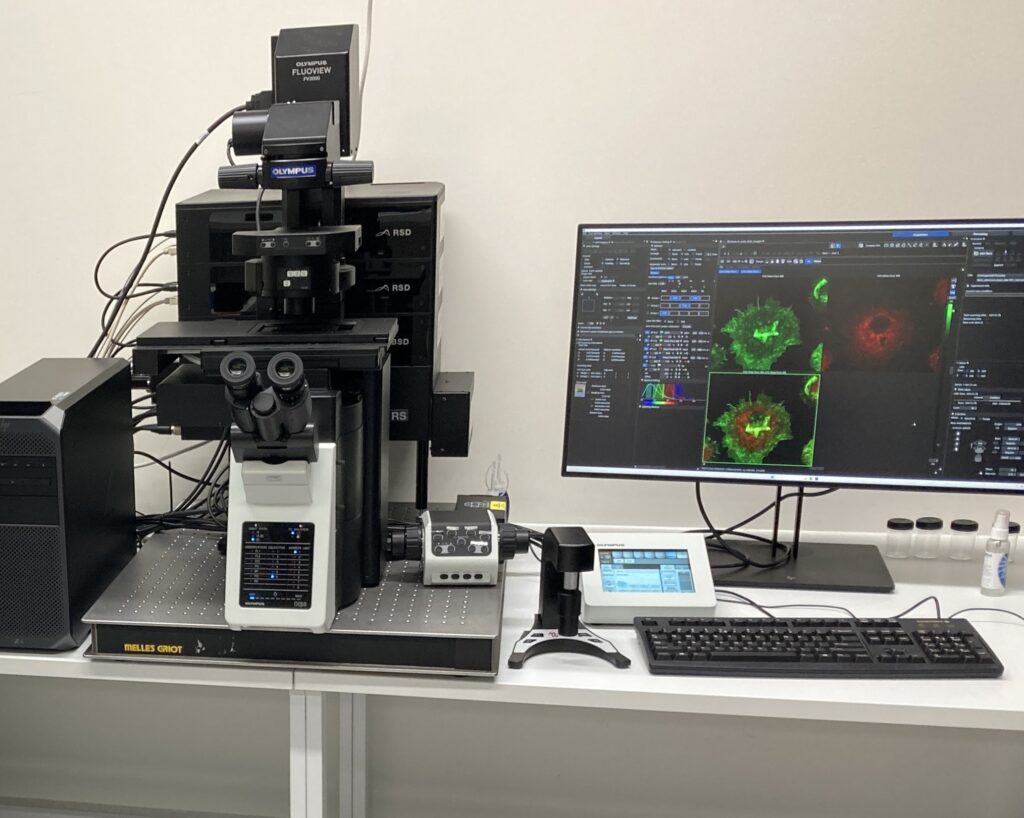
From February 5th to 9th 2024 Evident (formerly Olympus), in association with the OIC, will organize a demonstration of the newly released Fluoview FV4000 point scanning confocal.
Researchers who are interested to test the possibilities of this new generation laser scanning confocal are invited to bring their own samples for testing.
The FV4000 can image most of the regular samples used on the current OIC confocals. In addition, the confocal setup should be especially interesting for researchers using near infrared (NIR) fluorescent probes, or more than 4 fluorescent stains.
Special objectives will be available for imaging organoids and tissues. Specifications of the demonstration setup include:
Inverted microscope
10 laser spanning the wavelength range from 405 nm – to 785 nm
6 detectors for simulaneous imaging and spectral unmixing
Incubation system for live cell imaging (37°C/5% CO2) using 35mm dishes, multiwell slides and multiwell dishes
Resonant scanner for fast (>20fps) imaging
If you are interested to test the FV4000, please send us an email with a short description of the sample you would like to image.
In december the new Zeiss LSM900 is in Be-311 will be available for use. After the installation this summer we have fully tested and optimized the system for our imaging facility.
About the microscope
This microscope is also for fixed specimens, has 3 detectors and has the Zen-blue 3.7 software with an extended tile-scan module.
LSM700 users who did the introduction course can have an extra short (1 hour) introduction on the new microscope, mostly to show the differences and extended possibilities compared to the lsm700. Extra manuals will be provided via our website. Both the LSM700 systems will remain in our imaging facility.