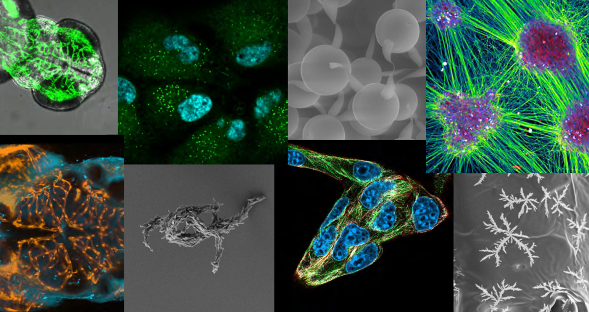
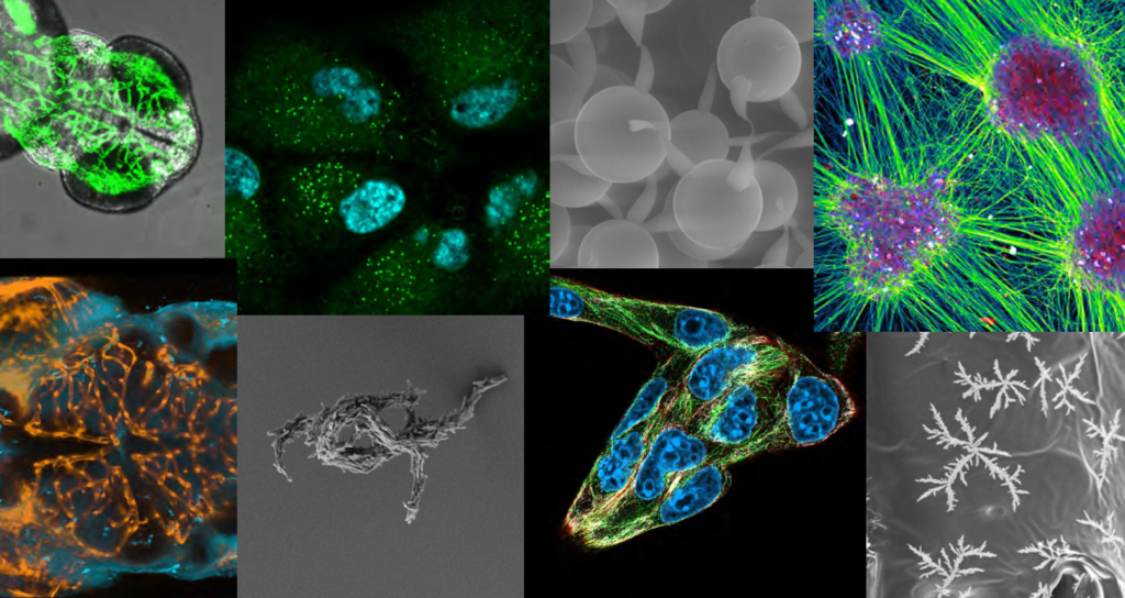
Submit your pictures for the Dutch Microscopy Pictures Calendar 2027. Dig out your best images, spread the word among your colleagues and networks, and feel free to think out of the box!
Deadline to submit: 15 September 2026 The best pictures will be featured on the 2027 Microscopy.nl birthday calendar. For questions email nl-bi@amsterdamumc.nl
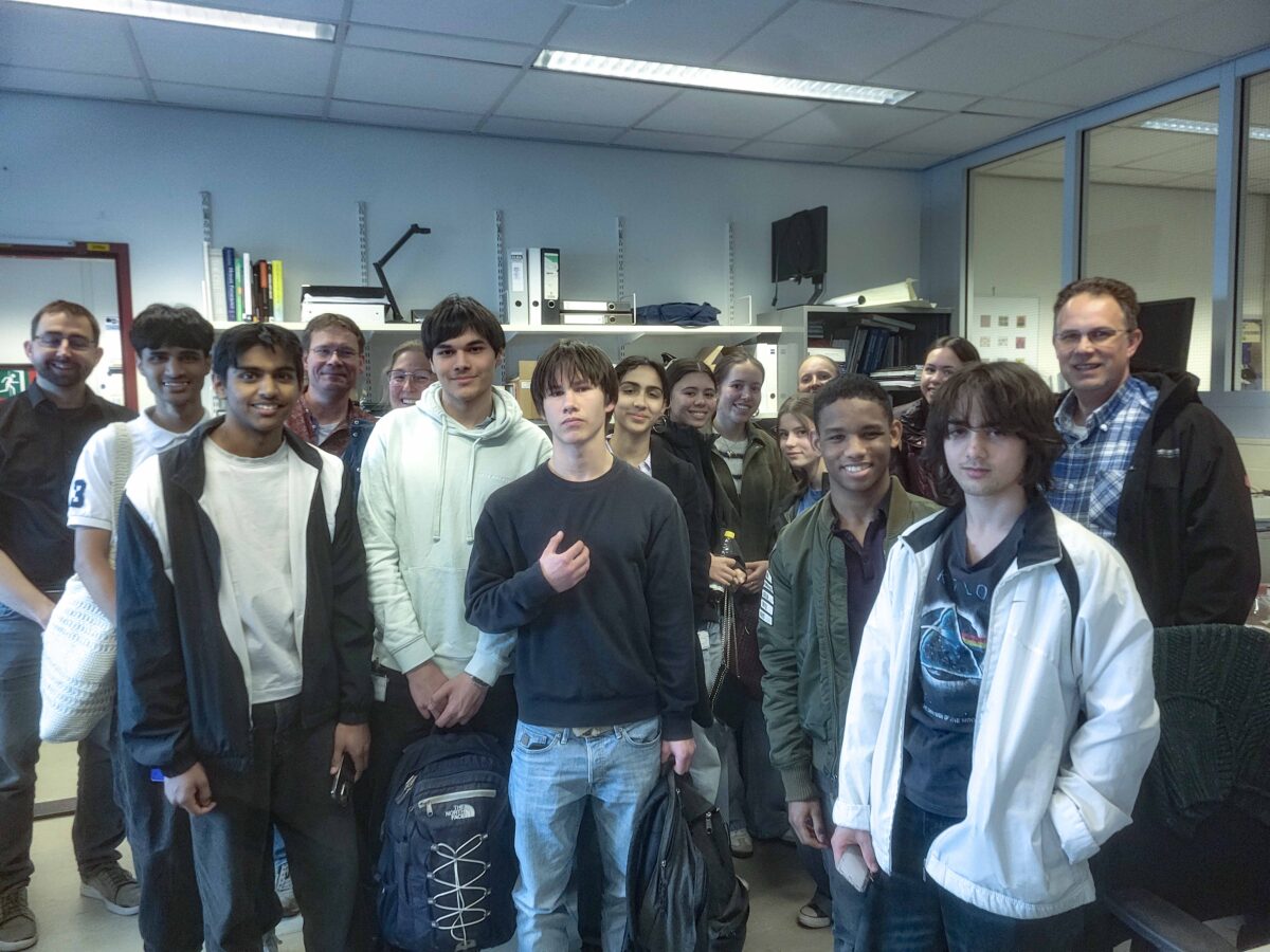
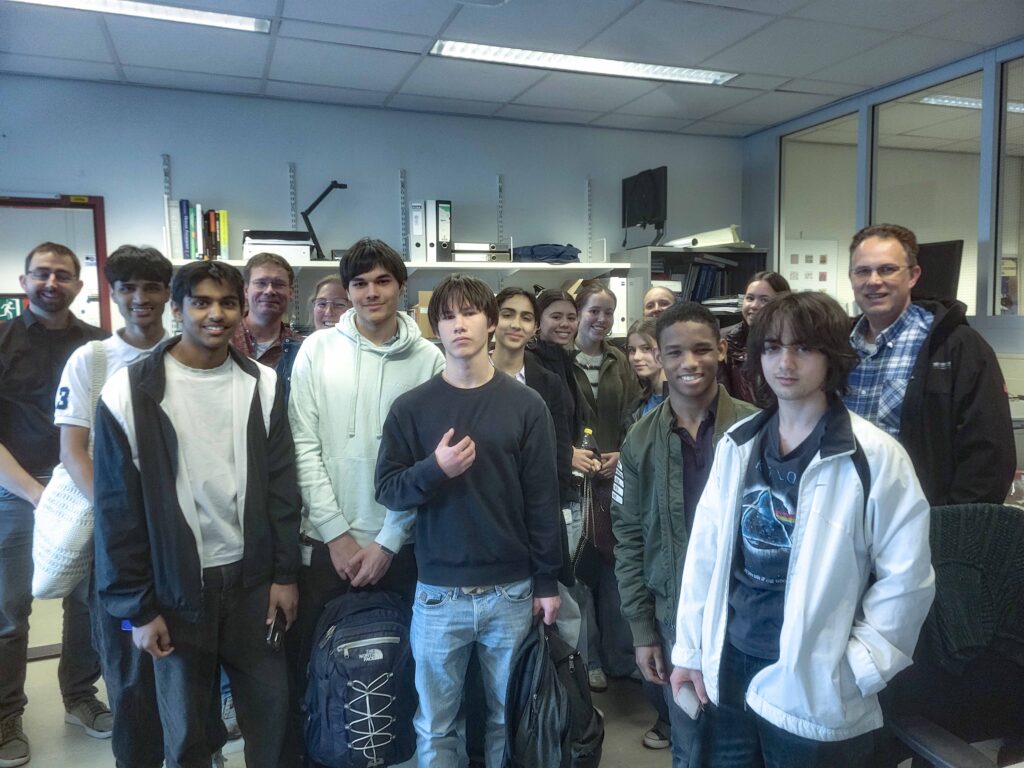
Onlangs ontving het Erasmus OIC een groep leerlingen van de Beta+ klas van het Emmaus College uit Rotterdam. Vooraf hadden zij op school al een gastles gekregen van OIC-ers Gert-Jan Kremers en Jeffrey van Haren over fluorescentie en confocale microscopie. Een mooie voorbereiding op wat ze tijdens hun bezoek in de praktijk konden ervaren.
Tijdens het bezoek maakten de leerlingen kennis met drie geavanceerde microscopietechnieken:
3D Switching: Met de confocale microscoop kan informatie uit verschillende diepten in lichaamscellen of weefsels worden gecombineerd tot een gedetailleerd driedimensionaal beeld. Dit geeft onderzoekers de mogelijkheid om complexe structuren beter te begrijpen. De studenten konden zelf met een laser in de cel fluorescente eiwitten van kleur veranderen en die dan in de tijd volgen.
Lightsheet microscopie: Een innovatieve techniek waarbij een zeer dunne laag (4 micrometer) licht wordt gebruikt om een stukje weefsel laagje voor laagje te belichten. Dit maakt het mogelijk om zeer snel grote biologische structuren levend in beeld te brengen. Een kloppend hartje van een zebravis-embryo kon zo gefilmd worden. Daarna werd het filmpje vertraagd afgespeeld om het rondgaan van de rode bloedcellen te volgen.
SIM Super Resolutie Microscopie: Structured Illumination Microscopy (SIM) doorbreekt de klassieke resolutiegrens van lichtmicroscopen. Hierdoor kunnen veel kleinere details zichtbaar worden gemaakt dan met standaardtechnieken mogelijk is. In levende cellen konden de kleine structuren van Focal Adhesions, de ‘voetjes van de cel’, beter in beeld gebracht worden.
Bij elke opstelling kregen zij niet alleen uitleg en zagen ze hoe de technieken in de praktijk werken, maar konden ze ook zelf met de microscopen aan de slag en zelf beelden maken.
Wij kijken terug op een geslaagd en leerzaam bezoek.
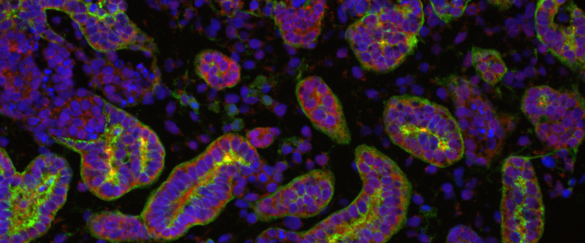
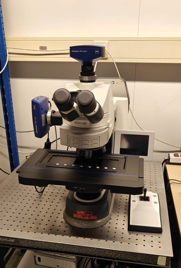
With this news post we would like to introduce the Zeiss AxioImager II Wide-Field microscope which has recently become part of our imaging centre.
This fully motorized system enables fast and scalable overview acquisition of fluorescence images in Wide-Field mode. Compatible with known channels across the visible spectrum and even into the near-infrared range.
An additional color camera allows you to correlate fluorescence images with a brightfield stain such as H&E. The 8‑slide insert makes it possible to process multiple samples in one run, significantly improving throughput for larger experiments.
Previously, this system was part of Pathology’s Research Facility (PARTS). We are pleased to make this microscope available once again within the Optical Imaging Centre. In the near future a software upgrade will make this system ready to be used for SMART microscopy. We will inform you in more detail when this is implemented.
Convenient pre-scan before acquiring confocal images
The microscope is equipped with a motorized stage capable of holding up to eight slides, thereby enabling the setup of large tile scans. The objectives are all air-immersion which makes it possible to adjust the magnification of the objective with the lowest magnification (2.5x) to the highest (63x) magnification without touching your sample.
Although widefield imaging provides lower z‑resolution than confocal microscopy, the system excels in scalability, acquisition speed and low magnification overview imaging. Combining the the two systems will give you an advantage in overview with the widefield and local detailed imaging with the confocal microscope.
The ability to transfer the overview images acquired with the Zeiss AxioImager II microscope to the Zeiss LSM 900 confocal system, also located in Be‑311, and subsequently acquire high‑resolution images of relevant regions, will be of significant value.
More specifications
The extra color camera will make this our only system in the imaging facility to correlate a chromogenic staining like H&E with fluorescence. Additional optics enable polarized light transmission imaging, expanding the range of contrast methods.
The Colibri 7 LED excitation system provides dedicated LEDs for each fluorescence channel, combining long lifetime with rapid switching. Its high transmission in the 735/40 nm band enables efficient near‑infrared imaging with reduced exposure times compared to other systems.
If you want to make use of this microscope, please contact us for an introduction. The system can be found in our schedular under the name WF_311.
More detailed specifications of this system can be found on our equipment page: link.
On Thursday and Friday February 5 & 6 2026 the OIC organizes, in cooperation with the Erasmus MC Graduate School a two-day Image analysis course “Microscopic Image Analysis: From Theory to Practice”. This course focuses on first time users and explains the basis of image analysis and how to use this in Fiji (ImageJ).
Course description
Microscopic images contain much more information than normally is retrieved. Extracting this information by visual inspection and manual measurement is not only cumbersome but also subjective. Automation of image analysis tasks by using a computer and the right software tools allows for higher efficiency, accuracy, objectivity, reproducibility, and completeness.
Requirements
Participants should bring their own laptop to the course for the practical exercises. Software and sample image data will be provided during the course. No prior knowledge of image processing is required. Participants have the opportunity to bring their own image data and directly apply the newly acquired image analysis techniques.
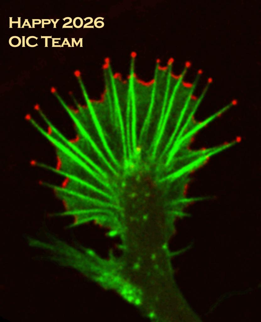
The Erasmus Optical Imaging Centre wishes you a happy 2026!!!
Tsion, Martijn, Marla, Johan, Jeffrey, Ilse, Gert-Jan, Bart, Alex and Adriaan
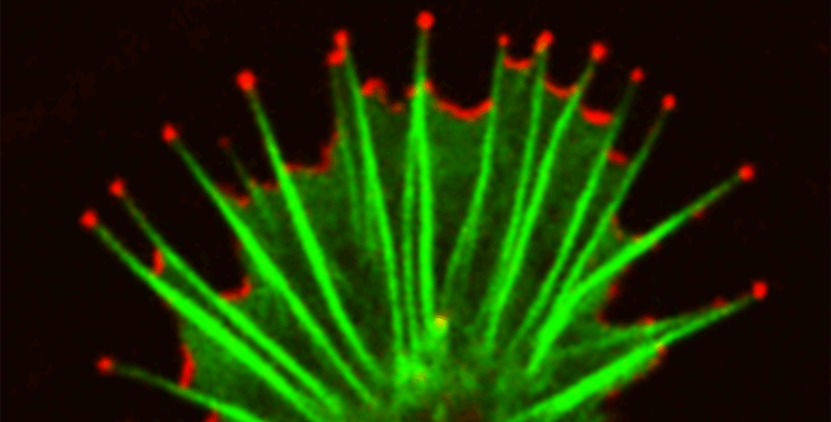
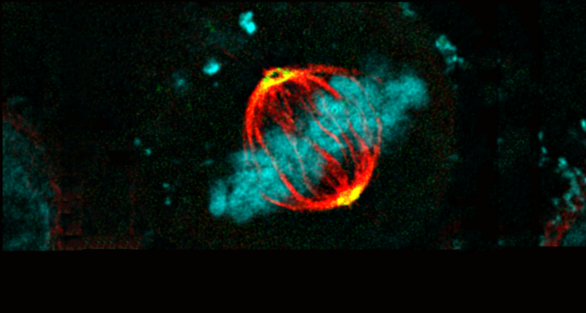
Confocal image of a live neuronal growth cone searching for a fellow neuron to make synapses together. Regulatory protein ABI1 (red) is present at the tips of growing actin filaments (green) as well as at the front edges of the lamellae connecting the filaments. Image recorded by Tim Allertz & Jeffrey van Haren, OIC.
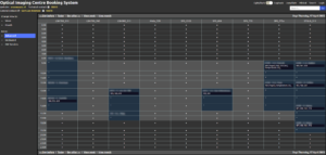
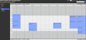
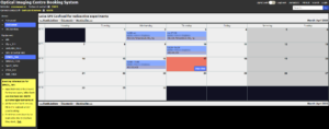
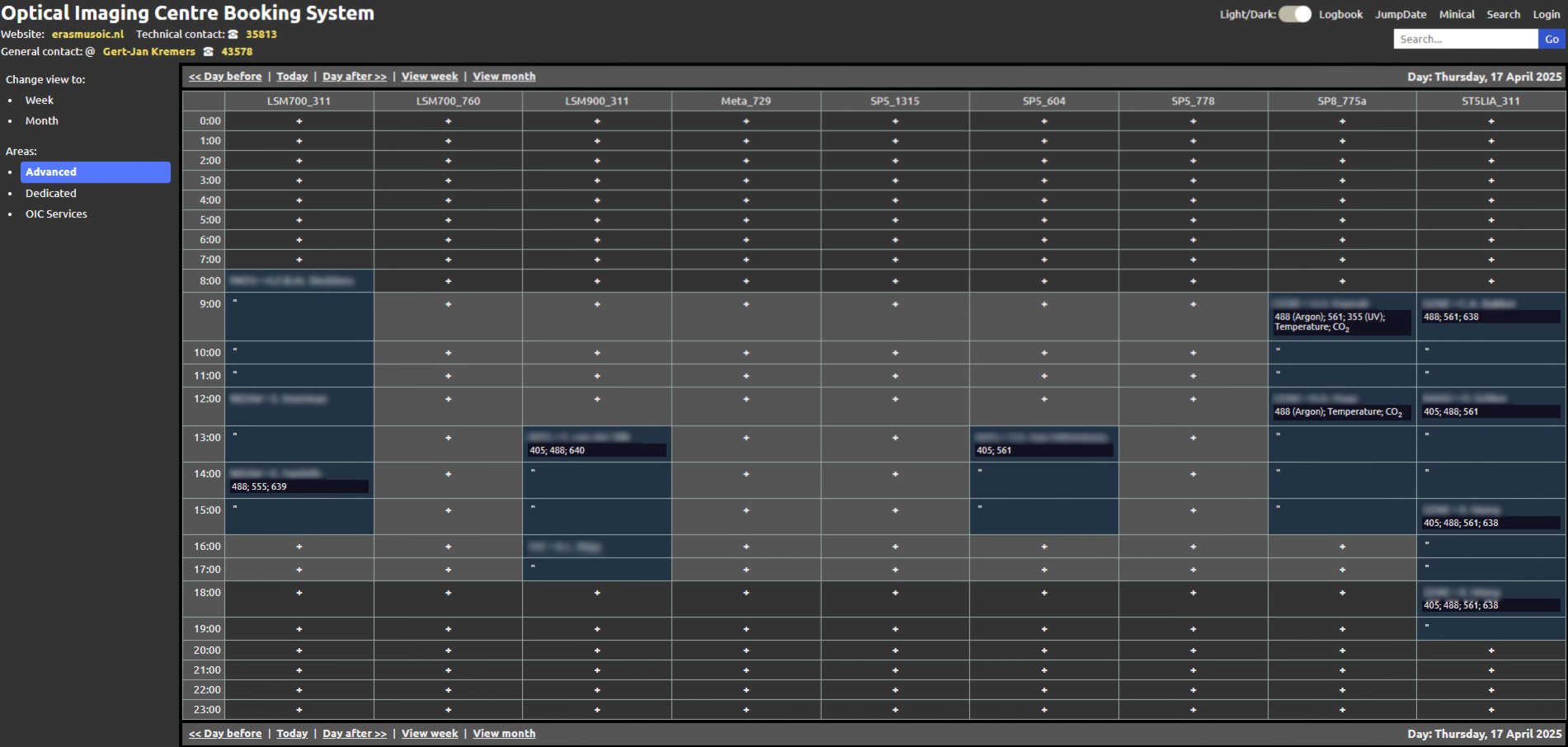
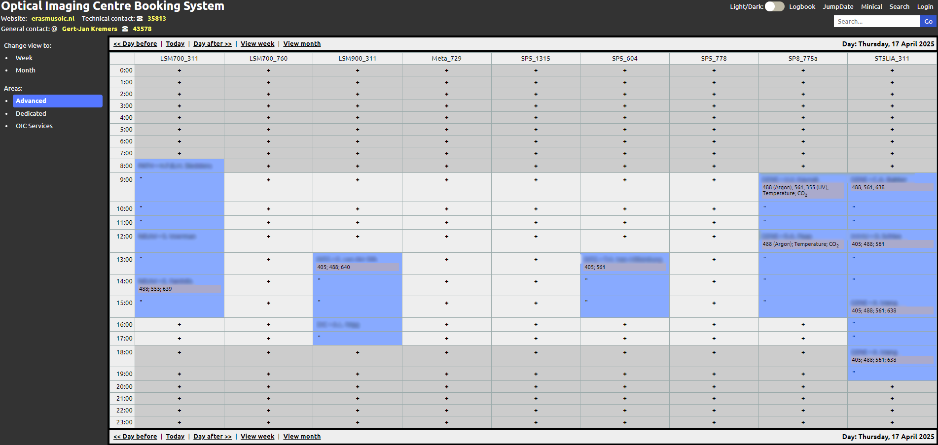
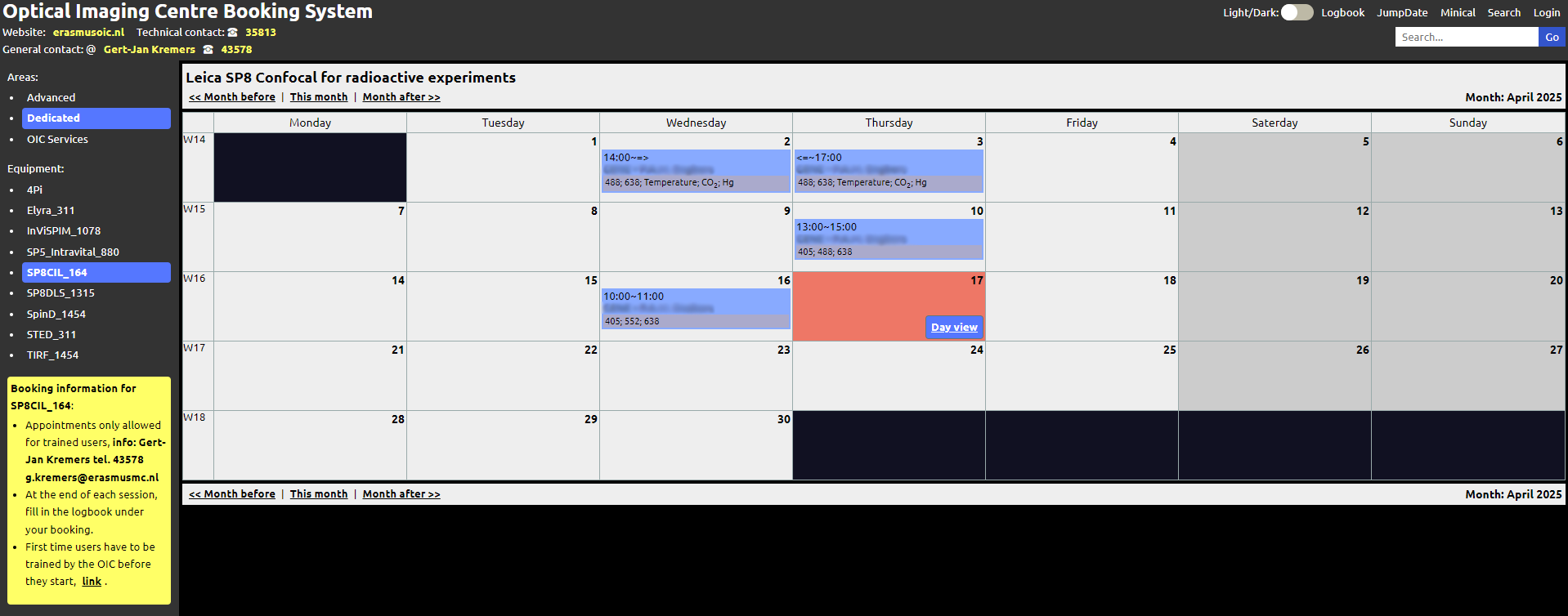
The microscope booking calendar has been updated with a few helpful new features. These changes make the interface more flexible and easier to use.
The calendar now supports both light and dark mode, with colors adjusted for clear visibility in each. In the month view, a new “Day” button allows users to quickly switch to the detailed daily view.
If you have any questions or feedback, feel free to reach out to the Erasmus OIC team.
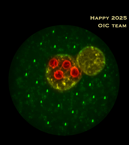
Confocal image (circular crop) of a fertilized mouse oocyte, with male and female pronuclei (yellow) and four prominent nucleoli (red) in the female pronucleus. The oocyte was decorated with ‘candle lights’ which are copies from the rightmost bright tiny structure in the cytoplasm (original image recorded by Gert van Cappellen, OIC)
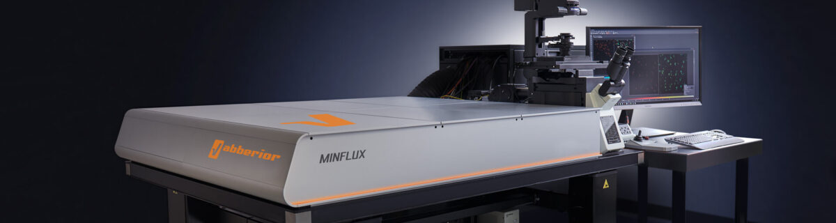
The Erasmus Optical Imaging Centre will host a MinFlux demonstration in the period of November 11-15 and a NVvM workshop focussed on MinFlux microscopy on Monday November 18.
MinFlux 3D unrivaled resolution and speed
MINFLUX, or minimal fluorescence photon fluxes microscopy, is a super-resolution light microscopy method that offers nanometer-scale 3D-imaging and microsecond-range single molecule tracking and can attain 1–3 nm resolution in three dimensions
We are excited to announce that our imaging facility has a state-of-the-art Abberior MinFlux system available for demonstation November 11-15. The system will also be part of the OIC functional imaging course in the same week. The demonstration will be concluded with a whole day NVvM MinFlux workshop on Monday November 18, 2024.
More information about the workshop, demonstration period and the registration can be found here.
We would like to introduce our new colleague Ilse Bakker, who started Juny of this year.
Ilse will be working in the OIC in the context of a project financed by the Convergence Flagship Imaging Facility and Innovation Centre (CIFIC) lead by Adriaan Houtsmuller, Erasmus MC and Jacob Hoogeboom, TU Delft. She will develop novel methods and staining techniques for combining fluorescent confocal microscopy and electron microscopy.
Registration is not longer possible for our PhD functional imaging and super-resolution course on November 11-15 2024.
The course is full, your registration will be placed on a waiting list. The course will be organized again in the fall of 2025.
This intensive course of one week gives an overview of the rapidly developing advanced fluorescence imaging methods and how they are used in biomedical research in a combination of lectures and hands-on practicals at our microscopes.
More information about the course and the registration form can be found via this link.